
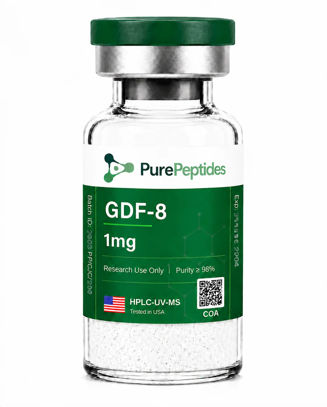
GDF-8 (Myostatin) Research Protein

$47.00
Supplied as a lyophilised powder and independently verified to ≥98% purity by HPLC and MS-UPLC analysis.
1000 in stock
🔥 Exclusive Crypto Offer
Get an INSTANT 15% DISCOUNT when you pay with Bitcoin, Ethereum, Dogecoin, Bitcoin Cash, XRP, Solana, or USDT.
⚡ Don't have crypto? Buy it here in 5 minutes with easy guidance.
💡 Pro Tip: You can also pay with cryptocurrency using your verified Cash App or PayPal account for a fast and secure checkout!
What is GDF-8 (Myostatin)?
GDF-8, better known as myostatin, is the natural protein that limits how much muscle the body builds. In research it is used as a reference standard and a tool, for example to test myostatin-blocking compounds or to model muscle loss in the lab.
- GDF-8 is the scientific name for myostatin, the natural brake on muscle growth
- Used in research as a reference standard for testing myostatin-pathway compounds
- Acts on ActRIIB receptors to limit muscle protein building
- Studied in animal models relevant to muscle loss
For research use only. Not approved for human therapeutic use.
GDF-8 (Growth Differentiation Factor 8), also known as Myostatin, is a recombinant protein belonging to the transforming growth factor-beta (TGF-beta) superfamily. The mature bioactive form exists as a disulfide-linked homodimer with an approximate molecular weight of 25,000 g/mol, comprising two 109-amino acid monomers. Each monomer contains the canonical seven-cysteine motif common to TGF-beta superfamily members. Produced via recombinant expression in E. coli, Myostatin signals through activin type II receptors (ActRIIA and ActRIIB) and is one of the most extensively studied negative regulators of skeletal muscle mass in preclinical research.
GDF-8 has been extensively investigated in preclinical musculoskeletal biology and metabolic research. Published in vitro studies in primary human myotube and C2C12 myoblast cell preparations have investigated GDF-8’s receptor binding characteristics at ActRIIB and downstream SMAD2/3 phosphorylation cascades, documenting myostatin-mediated inhibitory signalling and suppression of myoblast differentiation and myotube growth parameters under controlled experimental conditions [1]. The observation that targeted disruption of the GDF-8 gene in rodent models produces widespread increases in skeletal muscle mass has made Myostatin a key reference compound in studies examining muscle homeostasis, satellite cell quiescence, and the balance between anabolic and catabolic signalling within the TGF-beta pathway. Research has also examined GDF-8’s interaction with endogenous antagonists including Follistatin, with published rodent transgenic model studies documenting that Follistatin-mediated antagonism of myostatin activity produces widespread skeletal muscle increases consistent with those observed following targeted gene disruption, characterising the molecular determinants of ligand-antagonist engagement within the GDF-8 regulatory axis [2].
GDF-8 is produced to research-grade standards and independently verified by third-party HPLC and MS-UPLC analysis before dispatch. Vials are vacuum sealed and stored in a temperature controlled, monitored cold storage system. Certificates of Analysis are available on request.
Sold strictly for in vitro research purposes only. Not for human consumption. Intended for use by qualified researchers in laboratory settings only.
References
1Trendelenburg AU, Meyer A, Rohner D, Boyle J, Hatakeyama S, Glass DJ. Myostatin reduces Akt/TORC1/p70S6K signaling, inhibiting myoblast differentiation and myotube size. Am J Physiol Cell Physiol. 2009 Jun;296(6):C1258-1270. .PubMed PMID: 193572332Lee SJ, McPherron AC. Regulation of myostatin activity and muscle growth. Proc Natl Acad Sci U S A. 2001 Jul 31;98(16):9306–11. ; PubMed Central PMCID: PMC55416.PubMed PMID: 11459935
Scientific Review

Dr. Martina Rossi, PhD
Scientific Contributor and Reviewer
Reviewed for scientific accuracy, 14 June 2026
View credentials →
GDF-8 (Growth Differentiation Factor 8), also known as Myostatin, is a recombinant protein belonging to the transforming growth factor-beta (TGF-beta) superfamily. The mature bioactive form exists as a disulfide-linked homodimer with an approximate molecular weight of 25,000 g/mol, comprising two 109-amino acid monomers. Each monomer contains the canonical seven-cysteine motif common to TGF-beta superfamily members. Produced via recombinant expression in E. coli, Myostatin signals through activin type II receptors (ActRIIA and ActRIIB) and is one of the most extensively studied negative regulators of skeletal muscle mass in preclinical research.
GDF-8 has been extensively investigated in preclinical musculoskeletal biology and metabolic research. Published in vitro studies in primary human myotube and C2C12 myoblast cell preparations have investigated GDF-8’s receptor binding characteristics at ActRIIB and downstream SMAD2/3 phosphorylation cascades, documenting myostatin-mediated inhibitory signalling and suppression of myoblast differentiation and myotube growth parameters under controlled experimental conditions [1]. The observation that targeted disruption of the GDF-8 gene in rodent models produces widespread increases in skeletal muscle mass has made Myostatin a key reference compound in studies examining muscle homeostasis, satellite cell quiescence, and the balance between anabolic and catabolic signalling within the TGF-beta pathway. Research has also examined GDF-8’s interaction with endogenous antagonists including Follistatin, with published rodent transgenic model studies documenting that Follistatin-mediated antagonism of myostatin activity produces widespread skeletal muscle increases consistent with those observed following targeted gene disruption, characterising the molecular determinants of ligand-antagonist engagement within the GDF-8 regulatory axis [2].
GDF-8 is produced to research-grade standards and independently verified by third-party HPLC and MS-UPLC analysis before dispatch. Vials are vacuum sealed and stored in a temperature controlled, monitored cold storage system. Certificates of Analysis are available on request.
Sold strictly for in vitro research purposes only. Not for human consumption. Intended for use by qualified researchers in laboratory settings only.
References
1Trendelenburg AU, Meyer A, Rohner D, Boyle J, Hatakeyama S, Glass DJ. Myostatin reduces Akt/TORC1/p70S6K signaling, inhibiting myoblast differentiation and myotube size. Am J Physiol Cell Physiol. 2009 Jun;296(6):C1258-1270. .PubMed PMID: 193572332Lee SJ, McPherron AC. Regulation of myostatin activity and muscle growth. Proc Natl Acad Sci U S A. 2001 Jul 31;98(16):9306–11. ; PubMed Central PMCID: PMC55416.PubMed PMID: 11459935
Scientific Review
Dr. Martina Rossi, PhD
Scientific Contributor and Reviewer
Reviewed for scientific accuracy, 14 June 2026
View credentials →CAS Number152918-18-8Molecular Weight~25,000 g/mol (mature monomer); ~52,000 g/mol (active dimer)Purity≥98%Physical FormLyophilised PowderManufacturingManufactured in an ISO9001 Certified LaboratoryTestingHPLC + MS-UPLCSKUGDF8
Lyophilised powder: store at -20 °C or below, away from light and moisture. Once reconstituted in an appropriate laboratory diluent (e.g. sterile water, PBS, or assay buffer), store at 2–8 °C and use within the validated period for your protocol. Do not refreeze.
GDF-8 (Growth Differentiation Factor 8), also known as Myostatin, is a recombinant protein belonging to the transforming growth factor-beta (TGF-beta) superfamily. The mature bioactive form exists as a disulfide-linked homodimer with an approximate molecular weight of 25,000 g/mol, comprising two 109-amino acid monomers. Each monomer contains the canonical seven-cysteine motif common to TGF-beta superfamily members. Produced via recombinant expression in E. coli, Myostatin signals through activin type II receptors (ActRIIA and ActRIIB) and is one of the most extensively studied negative regulators of skeletal muscle mass in preclinical research.
GDF-8 has been extensively investigated in preclinical musculoskeletal biology and metabolic research. Published in vitro studies in primary human myotube and C2C12 myoblast cell preparations have investigated GDF-8’s receptor binding characteristics at ActRIIB and downstream SMAD2/3 phosphorylation cascades, documenting myostatin-mediated inhibitory signalling and suppression of myoblast differentiation and myotube growth parameters under controlled experimental conditions [1]. The observation that targeted disruption of the GDF-8 gene in rodent models produces widespread increases in skeletal muscle mass has made Myostatin a key reference compound in studies examining muscle homeostasis, satellite cell quiescence, and the balance between anabolic and catabolic signalling within the TGF-beta pathway. Research has also examined GDF-8’s interaction with endogenous antagonists including Follistatin, with published rodent transgenic model studies documenting that Follistatin-mediated antagonism of myostatin activity produces widespread skeletal muscle increases consistent with those observed following targeted gene disruption, characterising the molecular determinants of ligand-antagonist engagement within the GDF-8 regulatory axis [2].
GDF-8 is produced to research-grade standards and independently verified by third-party HPLC and MS-UPLC analysis before dispatch. Vials are vacuum sealed and stored in a temperature controlled, monitored cold storage system. Certificates of Analysis are available on request.
Sold strictly for in vitro research purposes only. Not for human consumption. Intended for use by qualified researchers in laboratory settings only.
References
1Trendelenburg AU, Meyer A, Rohner D, Boyle J, Hatakeyama S, Glass DJ. Myostatin reduces Akt/TORC1/p70S6K signaling, inhibiting myoblast differentiation and myotube size. Am J Physiol Cell Physiol. 2009 Jun;296(6):C1258-1270. .PubMed PMID: 193572332Lee SJ, McPherron AC. Regulation of myostatin activity and muscle growth. Proc Natl Acad Sci U S A. 2001 Jul 31;98(16):9306–11. ; PubMed Central PMCID: PMC55416.PubMed PMID: 11459935
Scientific Review
Dr. Martina Rossi, PhD
Scientific Contributor and Reviewer
Reviewed for scientific accuracy, 14 June 2026
View credentials →CAS Number152918-18-8Molecular Weight~25,000 g/mol (mature monomer); ~52,000 g/mol (active dimer)Purity≥98%Physical FormLyophilised PowderManufacturingManufactured in an ISO9001 Certified LaboratoryTestingHPLC + MS-UPLCSKUGDF8
Lyophilised powder: store at -20 °C or below, away from light and moisture. Once reconstituted in an appropriate laboratory diluent (e.g. sterile water, PBS, or assay buffer), store at 2–8 °C and use within the validated period for your protocol. Do not refreeze.
Related products
GH Axis Research

GH Axis Research

GH Axis Research
GH Axis Research

GH Axis Research

GH Axis Research

GH Axis Research
GH Axis Research